![]()
![]()
![]()
![]()

|
|
|
|
Materials and Supplies
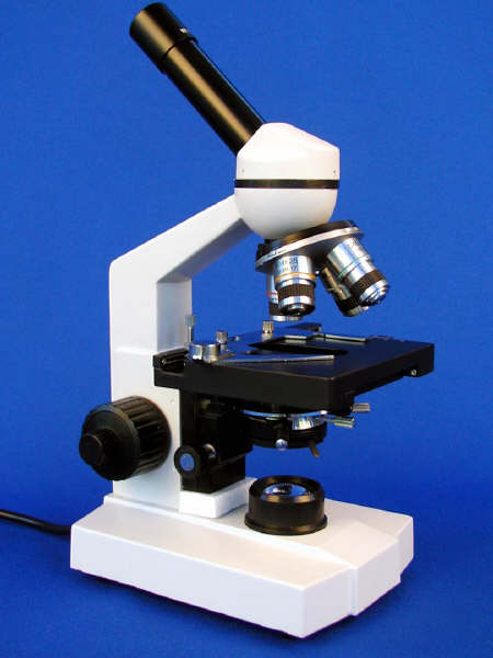
Floatation Fecal Analysis Method 1. Mix up the flotation solution. You can use a variety of chemicals including salt or sugar but my preference is sugar. Sugar solution is prepared by dissolving a pound (454 grams) of sugar in 1 4/5 cups (355 ml) of water, and salt takes a pound (454 grams) of salt in 4 4/5 cups (1140ml ) of water. Bring briefly to a boil to sterilize the solution. This can be done in a quart canning jar. Specific gravity should be approximately 1.27. 2. Collect fresh feces. Use an old pill bottle or a small jar for each animal. If not examined immediately, be sure to label the container with the date, time and animal that provided the specimen and keep it in a cool place. 3. Place 3 or 4 fresh beans into a small glass container (a baby food jar will do) and pour in some flotation solution to partially cover them (about a full test tube by volume). 4. Mash them up in the liquid with your stirring rod. Pour it through the strainer to remove the large particles into a second small glass container. Pour the strained liquid into a clean test tube. 5. If necessary, fill up the test tube to the very top with more liquid. Place a microscope coverslip over the top. There should be no air between the coverslip and the liquid. Over time (20-30 minutes) the eggs will float up to the top and adhere to the glass plate. If a centrifuge is available, this time can be reduced to five minutes. 6. Carefully remove the coverslip and lower it at an angle over a microscope slide with the sample sandwiched between both pieces of glass. 7. Examine the specimen for worm eggs and coccidia oocysts. Start with the lowest power (40X) on your microscope and increase magnification if you see something interesting. An illustrated chart would be helpful in identifying them. Note, you will also be looking at other debris. Do not confuse it with parasites. Modified Stoll's Fecal Analysis Method This method is more accurate than the simple
floatation method, but does require a centrifuge (preferably a swinging bucket
style).
1. 5 grams of feces is diluted with a measured quantity of regular tap water to a final quantity of 100 grams: 5 grams feces plus 95 ml water. 2. The fecal sample is dispersed evenly in the water, then 10 ml of the fecal slurry is decanted into a graduated centrifuge tube. This represents 10% of the original fecal sample (0.5 grams). 3. This sample is pelleted by centrifugation, the supernatant discarded, then the flotation solution is added to the centrifuge tube and mixed thoroughly. The tube is placed into the centrifuge, a slightly bulging meniscus is formed by adding several drops of additional flotation solution to the tube, and a coverslip is secured onto the meniscus and centrifuge tube. Centrifuge for 10 minutes at approximately 2000rpm. 4. Remove the coverslip after centrifugation and count all strongyle-type eggs that are recovered. We count only the strongyle-type eggs and make a notation if other parasitic structures are present. Based on this count of all strongyle-type eggs present in 10% of the original fecal sample, we establish the EPG by a simple multiplication constant of 2.
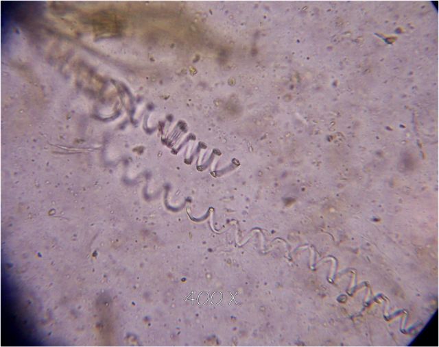
Video of live strongyloides (short - 294k)
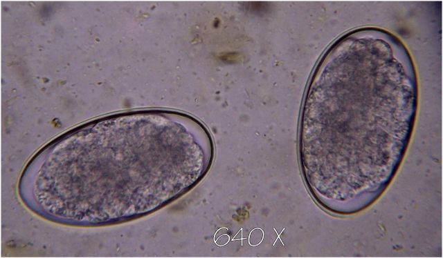
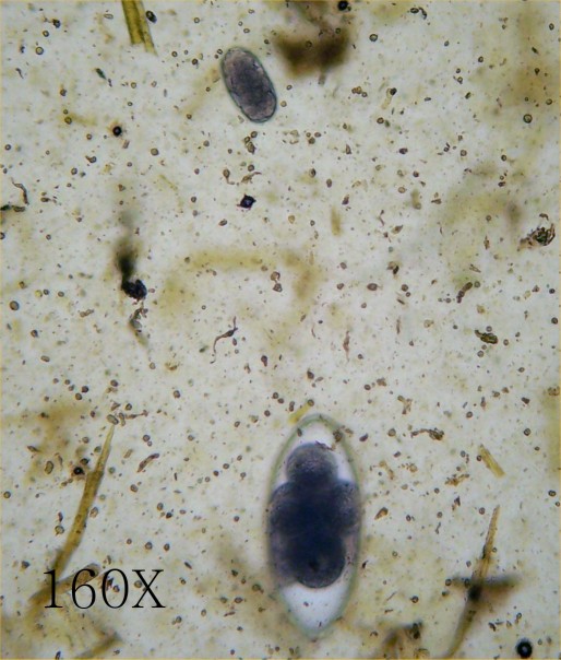
Strongyle oocyst (top) and Nematodirus oocyst (bottom)
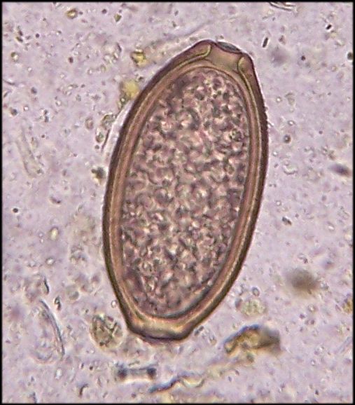
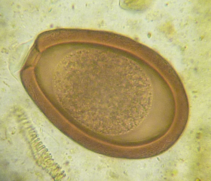
Taenia (tapeworm) egg.
Here's one along with a Nematodirus for reference. Note the much larger than usual tapeworm egg size.
Capillaria. One egg found in llama.
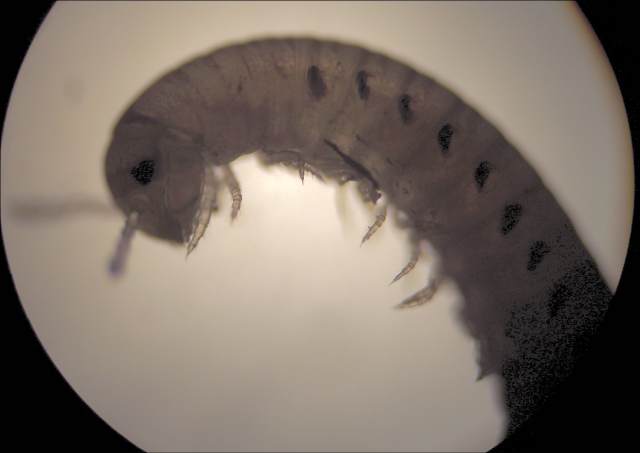
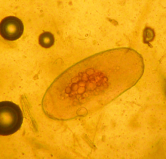
Unidentified worm found in feces (different llama) after treatment with fenbendazole. Length is about a half inch. Not what I would expect from an intestinal worm. What would intestinal parasites do with antenna?
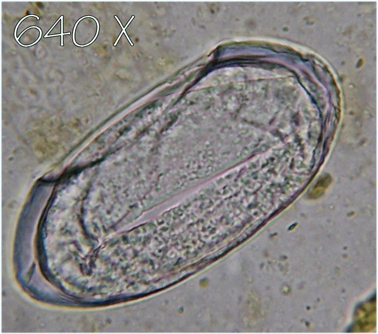
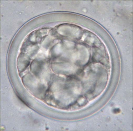
The size of a Nematodirus, and with a few Nematodirus eggs found on the slide, this one has a very different nucleus both in structure and color. There was only one. Malformed Nematodirus?
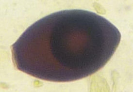
Coccidia, Emeria macusaniensis (E. mac)
Coccidia, Emeria, specific species unknown. It is a small fraction of the size of E. mac. -- only a speck at at low magnification. |
|
Send mail to
RRR with
questions or comments about this web site.
|